Table of Contents >> Show >> Hide
- What a “glaucoma test” actually means
- Purpose of glaucoma testing
- Types of glaucoma tests (and what each one tells your doctor)
- What to expect at a glaucoma testing appointment
- How doctors interpret results (without turning you into a statistic)
- FAQ: Glaucoma testing questions people actually ask
- Does a glaucoma test hurt?
- Can you diagnose glaucoma from eye pressure alone?
- How often should I get glaucoma testing?
- Do I need someone to drive me home after dilation?
- What if my visual field test is “unreliable”?
- Can glaucoma tests detect early disease?
- Are glaucoma tests covered by insurance or Medicare?
- What’s the difference between an optometrist and an ophthalmologist for glaucoma testing?
- When you should consider scheduling a glaucoma evaluation
- Experiences: What glaucoma testing feels like in real life (the human side)
- The “air puff betrayal” moment
- The “why is the waiting room suddenly so bright?” dilation phase
- The visual field test: equal parts focus and self-doubt
- OCT imaging: the easiest “hard” test you’ll ever do
- Gonioscopy: sounds scary, feels mostly odd
- The emotional part: relief, worry, and the power of a plan
- Practical tips patients wish they knew beforehand
- Conclusion
If you’ve ever had an eye appointment where a machine “puffs” air at your eyeball, you’ve already met one
member of the glaucoma-testing crew. Unfortunately, glaucoma isn’t the kind of villain that announces itself
with theme music. Early on, it can be symptom-free, quietly affecting the optic nerve and peripheral vision.
That’s why glaucoma testing is less like a single “gotcha” moment and more like a well-run investigation:
multiple clues, multiple tools, one goalcatch problems early and track them accurately over time.
This guide explains what glaucoma tests are, why you might need them, what each test measures, and the
most common questions people ask (including “Will this hurt?” and “Do I need a driver after dilation?”).
Spoiler: most tests are quick, many are painless, and none require you to be brave in the cinematic sense.
Just blink normally and try not to challenge the machine to a staring contest.
What a “glaucoma test” actually means
A “glaucoma test” usually isn’t one test. It’s a set of exams that help an eye care professional evaluate:
(1) pressure inside the eye (intraocular pressure, or IOP), (2) the optic nerve’s health, (3) the drainage angle
where fluid leaves the eye, and (4) whether your visual field (especially side vision) shows patterns consistent
with glaucoma.
This matters because glaucoma is not a one-metric disease. Some people develop optic nerve damage at
relatively “normal” pressures, while others can tolerate higher pressures without immediate damage.
So eye pressure is importantbut it’s not the entire story. Think of IOP like the speedometer: useful, but you
still need the windshield, mirrors, and the rest of the dashboard to drive safely.
Purpose of glaucoma testing
1) Screening: catching risk before vision is affected
Many people pursue testing because they’re at higher risk: age, family history, certain ancestry backgrounds,
diabetes, thinner corneas, higher eye pressure, or previous suspicious findings on routine exams.
Screening aims to identify early changes so treatment can start soonerbecause vision lost to glaucoma is
generally not reversible.
2) Diagnosis: confirming whether glaucoma is present
Diagnosis typically relies on a combination of findings: optic nerve appearance, imaging changes, visual field
results, IOP patterns, and angle assessment. One isolated result rarely “proves” glaucoma on its own. The more
consistent the pattern across tests, the clearer the diagnosis.
3) Monitoring: tracking progression and treatment response
If you already have glaucoma (or you’re a “glaucoma suspect”), repeat testing helps determine whether the
condition is stable or progressing. Monitoring often includes periodic visual field testing and optic nerve imaging,
plus IOP measurement at visits. The goal is to keep you seeing well for the long haul.
Types of glaucoma tests (and what each one tells your doctor)
Tonometry (eye pressure test)
Tonometry measures intraocular pressure (IOP). You might experience the “air puff” version or the
numbing-drop version where a tiny probe gently touches the eye’s surface (often called applanation tonometry).
The test is fast and usually feels like… almost nothing, aside from the surprise factor of the puff.
Important nuance: IOP is a key risk factor, but pressure alone can’t confirm or rule out glaucoma. Your cornea’s
thickness and other factors can influence readings, and some glaucoma occurs with pressures in the average
range. That’s why tonometry is typically paired with other exams.
Pachymetry (corneal thickness measurement)
Pachymetry measures central corneal thickness. Why should you care about corneal thickness? Because it helps
interpret IOP readings. A thinner or thicker cornea can affect how pressure measurements read, and corneal
thickness may also help assess overall risk in certain situations.
Pachymetry is quick and commonly painless. Depending on the device, you may have a numbing drop, and a
small instrument may lightly touch the eyeor it may be measured without contact.
Gonioscopy (drainage angle exam)
Gonioscopy evaluates the angle where the iris meets the corneathe eye’s internal “drain” area where fluid
typically exits. This helps distinguish open-angle vs. angle-closure mechanisms and guides treatment decisions.
The exam uses a special lens placed gently on the eye after numbing drops. It sounds intense, but it’s usually
more “weird” than painfullike wearing a tiny contact lens you didn’t ask for.
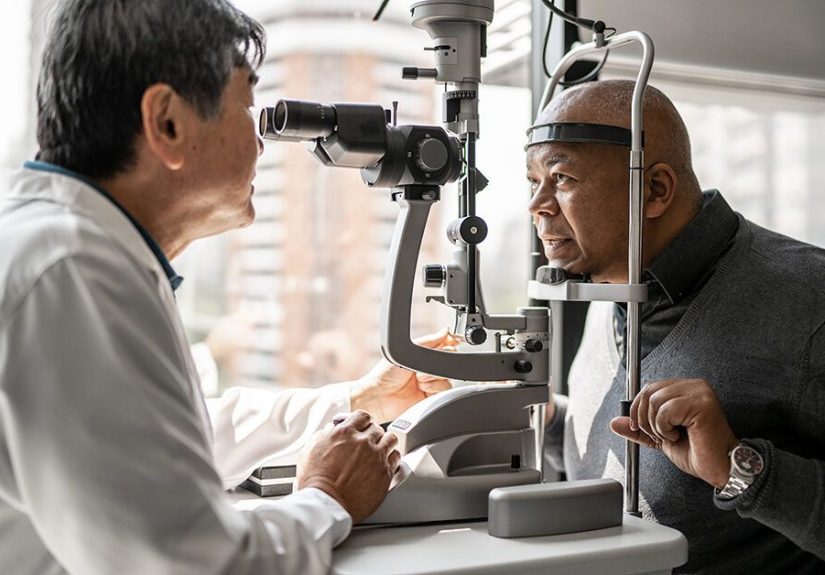
Dilated eye exam / ophthalmoscopy (optic nerve evaluation)
With dilation drops, your pupils widen so the clinician can get a clearer view of the optic nerve and retina.
During this exam, they look for changes in the optic nerve head (such as “cupping”) and other findings that may
suggest glaucoma or another condition.
Dilation can cause blurry near vision and light sensitivity for a few hours. You’ll want sunglasses afterward, and
you may not love drivingespecially at nightuntil your vision feels normal again.
Visual field test (perimetry)
Perimetry maps your field of vision, with special attention to side (peripheral) visionoften where glaucoma
changes show up first. You’ll stare at a central target inside a dome-like machine and press a button whenever
you see a light spot. The test sounds simple, but it requires focus, and it’s normal to feel uncertain about a few
flashes. The machine expects that. It’s not judging you.
Visual field tests are essential for detecting functional vision loss patterns and for monitoring progression over
time. If your first test is “messy,” don’t panic. There’s a learning curve, and repeat tests often become more
reliable as you get used to it.
Optic nerve imaging (OCT and photos)
Optical coherence tomography (OCT) provides detailed measurements of retinal nerve fiber layer and related
structuresareas that can thin with glaucoma damage. Many clinics also capture optic nerve photos to compare
changes over time. Imaging helps detect structural change, sometimes before noticeable vision symptoms appear.
Imaging is usually quick, noninvasive, and feels like taking a fancy eye selfieexcept you’re not allowed to pick
the filter.
Additional tests you might see (depending on your case)
- Slit-lamp exam: a microscope-based exam of the front structures of the eye.
- Anterior segment imaging: additional views of the drainage angle or front-of-eye anatomy.
- Repeat IOP checks: sometimes pressure varies by time of day, so patterns matter.
What to expect at a glaucoma testing appointment
A typical “glaucoma workup” may include a sequence like: vision check, pressure measurement, corneal thickness
measurement, dilation, optic nerve evaluation, imaging, and/or a visual field test. You might not do every test
every visitclinics often schedule visual fields and imaging at different intervals depending on risk and stability.
The whole visit can range from 30 minutes to a couple hours, mostly because dilation takes time and visual field
testing takes concentration. Pro tip: eat beforehand. Nothing makes “press the button when you see the light”
harder than being hangry.
How doctors interpret results (without turning you into a statistic)
They’re looking for patterns, not perfection
Glaucoma assessment is pattern recognition across multiple data points: optic nerve appearance, OCT thickness
trends, visual field defects, and risk factors. A single borderline reading may prompt repeat testing rather than an
immediate diagnosis.
Baseline matters more than “normal”
Many clinicians build a baseline: what your optic nerve looks like, how your visual field behaves, what your IOP
range is, and how thick your corneas are. Future tests are compared to younot just a generic reference range.
Progression is a big deal
Glaucoma care often focuses on whether there’s progression over time. Stable findings may mean the current plan
is working. Evidence of change may trigger treatment adjustments, additional testing, or closer follow-up.
FAQ: Glaucoma testing questions people actually ask
Does a glaucoma test hurt?
Most glaucoma tests are painless. Tonometry may feel like a gentle tap or a brief puff of air. Gonioscopy and
applanation tonometry typically use numbing drops. Visual field testing doesn’t touch your eye at allit just tests
your patience (politely).
Can you diagnose glaucoma from eye pressure alone?
No. Eye pressure is important, but it’s not enough by itself. Glaucoma can occur with “normal” pressures, and
elevated pressure doesn’t always mean glaucoma. That’s why comprehensive evaluation includes optic nerve
assessment, visual field testing, and often imaging and angle evaluation.
How often should I get glaucoma testing?
The schedule depends on your risk factors and findings. Some people need regular comprehensive dilated eye
exams even without symptoms, and those at higher risk may be advised to return more frequently. If you’re a
glaucoma suspect or diagnosed, your clinician will tailor follow-up intervals and which tests to repeat (for example,
visual fields and OCT at set intervals).
Do I need someone to drive me home after dilation?
Dilation can blur near vision and increase light sensitivity for a few hours. Many people can get home fine, but
drivingespecially at nightmay feel uncomfortable or unsafe. If you’re unsure, bring sunglasses and consider
arranging a ride, particularly if you know your eyes stay blurry for a while after drops.
What if my visual field test is “unreliable”?
It happens. Visual fields can be affected by learning curve, fatigue, dry eyes, blinking, distraction, or simply being
human. Clinicians often repeat tests to confirm patterns. Don’t interpret one imperfect test as a life sentence.
Treat it as a draft, not the final novel.
Can glaucoma tests detect early disease?
Yesespecially when multiple tools are used. Imaging (like OCT) can identify structural changes, while visual field
testing can detect functional loss patterns. Early detection is one of the main reasons routine eye exams are so
valuable, even when you feel fine.
Are glaucoma tests covered by insurance or Medicare?
Coverage varies by plan and medical necessity. Medicare has specific coverage provisions for glaucoma testing
for people in higher-risk groups (for example, screening frequency and eligibility rules). If cost is a concern, ask
the clinic what codes they’re billing and whether prior authorization is needed.
What’s the difference between an optometrist and an ophthalmologist for glaucoma testing?
Both can perform key glaucoma tests and detect suspicious findings. Ophthalmologists are medical doctors who
can provide surgical care and manage complex cases. Optometrists commonly provide ongoing eye care, testing,
and management depending on state scope of practice and the specifics of your case. What matters most is
timely evaluation and appropriate follow-up.
When you should consider scheduling a glaucoma evaluation
- If you have a family history of glaucoma
- If you’re older (risk increases with age)
- If you’ve been told you have elevated IOP or “large cup-to-disc ratio”
- If you have diabetes or other health conditions that increase eye disease risk
- If you notice peripheral vision changes (even subtle ones)
- If you’ve had eye injuries or long-term steroid use
If you have sudden eye pain, headache, halos, nausea, or rapid vision changes, treat that as urgent and seek
immediate caresome forms of angle-closure glaucoma can be an emergency.
Experiences: What glaucoma testing feels like in real life (the human side)
Medical descriptions are helpful, but they don’t always answer the real question people have in their heads:
“Okay… but what will it actually be like when I’m sitting in the chair?” Here are common experiences patients
report, bundled into realistic scenarios. (Not everyone’s visit is identical, but these patterns are very typical.)
The “air puff betrayal” moment
Many people remember their first air-puff tonometry test for one reason: surprise. You lean in, you try to keep
your eyes wide open like a heroic statue, and thenpfft. The most common reaction is not pain, but indignation:
“You could’ve warned me!” The good news: it’s over quickly, and it doesn’t leave lasting discomfort. The second
puff is usually easier because you stop trying to negotiate with the machine.
The “why is the waiting room suddenly so bright?” dilation phase
Dilation drops often feel mildly stingy for a few seconds. Then comes the waiting period, where you sit and your
pupils widen. People commonly describe a gradual shift: phones get blurrier up close, indoor lighting feels harsher,
and you become strangely interested in sunglasses as a lifestyle choice. Many clinics are used to this and will tell
you what’s normal. The most consistent “after” experience is light sensitivity and near blur for a few hours.
The visual field test: equal parts focus and self-doubt
Perimetry is the test that makes people question their own reality. The lights are faint. You’re not supposed to
move your eyes. You blink and worry you missed something. Then you press the button and wonder if you pressed
too late. This is normal. Most patients improve on the second or third attempt because they understand the rhythm:
relax your shoulders, blink normally, keep your gaze steady, and respond when you’re reasonably sure.
Clinicians interpret results with reliability indicators and patternsso one “off” test usually leads to a repeat, not
a dramatic announcement.
OCT imaging: the easiest “hard” test you’ll ever do
OCT and optic nerve photos are often the least stressful part. Patients usually describe it as “staring at a target
light while the machine takes pictures.” No pokes, no puffs, no button-mashing. The only challenge is holding
still and keeping your eyes open long enough for the scan to finish. Many people are surprised that such a quick
scan can provide meaningful information about nerve fiber layers and subtle change over time.
Gonioscopy: sounds scary, feels mostly odd
If you’re told you need gonioscopy, it can sound intense because it involves a lens touching the eye. In practice,
numbing drops do the heavy lifting. Patients most often describe a gentle pressure and a “contact lens” sensation.
The uncomfortable part is usually psychological: you’re aware something is near your eye, and your brain is trying
to protect you by blinking. Once you realize it doesn’t hurt, the exam tends to be manageable.
The emotional part: relief, worry, and the power of a plan
Glaucoma testing can bring up feelingsespecially if a family member had glaucoma or if you’re hearing words
like “suspect” or “monitor.” A common experience is a mix of relief (“I’m finally getting checked”) and worry
(“What if they find something?”). Many people feel better once they understand that glaucoma care is often a
long-term management strategy, not an immediate crisis: establish a baseline, follow trends, treat risk, and protect
vision. Even when results are abnormal, having a clear planmedication, follow-up intervals, repeat testingcan
reduce anxiety because uncertainty becomes a roadmap.
Practical tips patients wish they knew beforehand
- Bring sunglasses: dilation plus daylight can feel like staring at the sun’s resume.
- Plan your day: schedule detailed close-up work (and long drives) for after your vision clears.
- Rest before visual fields: fatigue can make the test harder than it needs to be.
- Ask what “baseline” means for you: understanding the plan can make follow-up feel purposeful.
- Don’t panic over one result: glaucoma testing is often about repeatability and trends.
Conclusion
Glaucoma testing is a team sport: eye pressure checks, optic nerve exams, imaging, visual field testing, corneal
thickness measurement, and angle evaluation all play different roles. The purpose is simple but powerfuldetect
risk early, diagnose accurately, and monitor for change so vision can be protected over time.
If you’re due for an eye exam or you have risk factors, don’t wait for symptoms to appear. Glaucoma often starts
quietly, but your testing doesn’t have to be scary. It can be straightforward, data-driven, and (with the right
mindset) only mildly annoyinglike most adult responsibilities.