Table of Contents >> Show >> Hide
- Quick Definitions (So We’re Speaking the Same Skin Language)
- Why SK and Melanoma Get Confused
- Seborrheic Keratosis: What It Usually Looks and Feels Like
- Melanoma: What to Watch For (ABCDE + “Ugly Duckling”)
- Seborrheic Keratosis vs. Melanoma: Side-by-Side Comparison
- Red Flags: When an “SK” Might Not Be “Just an SK”
- What to Expect at the Dermatologist
- Treatment Differences (Because the Plan Depends on the Diagnosis)
- Risk Factors and Prevention You Can Actually Use
- FAQs People Ask (Usually While Staring at a Spot in the Mirror)
- The Bottom Line: A Simple Rule That Covers 99% of Real Life
- Real-World Experiences: What People Commonly Go Through When Comparing SK vs. Melanoma
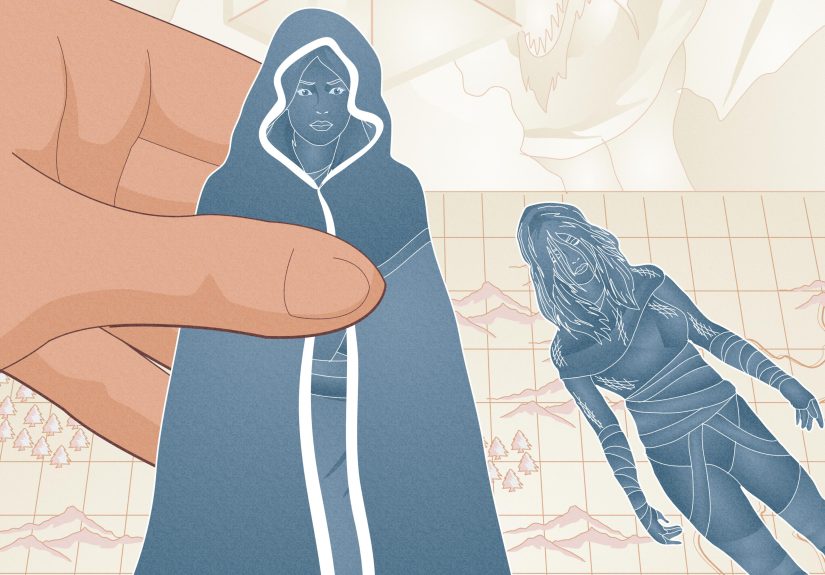
Your skin is basically your body’s billboardexcept you didn’t ask for the ads. One day you notice a new spot that looks like it was
“pasted on,” and your brain immediately jumps to: Is this skin cancer? Totally normal reaction.
Here’s the good news: seborrheic keratoses (often called “SKs”) are very common and usually harmless. The not-so-fun truth:
melanoma is a serious type of skin cancer that needs prompt attention. And to make things extra confusing, an SK can sometimes
look a little like melanoma (and vice versa).
This guide breaks down what seborrheic keratosis is, what melanoma is, how they typically look and behave, and when you should stop Googling
and let a dermatologist do their detective work. (Spoiler: a quick examand sometimes a biopsyis the real “know for sure” button.)
Important: This article is for education, not diagnosis. If a spot is new, changing, bleeding, or just looks “off,” get it checked.
Quick Definitions (So We’re Speaking the Same Skin Language)
What is seborrheic keratosis?
A seborrheic keratosis is a benign (noncancerous) skin growth that often appears as people get older. Many look waxy,
scaly, or warty, with that classic “stuck-on” appearancelike someone pressed a tiny piece of candle wax onto your skin.
SKs can show up as a single spot or in groups, and they commonly appear on the chest, back, face, scalp, neck, or shoulders.
What is melanoma?
Melanoma is a skin cancer that starts in pigment-producing cells (melanocytes). It often shows up as a changing mole,
a new suspicious spot, or a lesion that doesn’t follow the “rules” of your other moles. Melanoma can be highly treatable when caught early,
but it can become dangerous if it grows deeper or spreadsso early detection matters.
Why SK and Melanoma Get Confused
In a perfect world, every harmless spot would look obviously harmless and every dangerous spot would wear a tiny name tag that says,
“Hi, I’m melanoma.” Unfortunately, skin doesn’t work like that.
Some SKs can be very dark, irregular, crusty, or inflamed (especially if they rub on clothing). Some melanomas can be subtle, small,
or oddly shaped. And some melanomas don’t look “classic” at all (for example, they can appear pink or skin-colored).
That’s why dermatologists use pattern recognition, tools like dermoscopy, andwhen neededbiopsy. A photo, a friend’s opinion, or a
“spot-check app” can’t reliably replace that process.
Seborrheic Keratosis: What It Usually Looks and Feels Like
Seborrheic keratoses come in a variety packdifferent shades, sizes, and textures. Still, many share a few classic traits:
- “Stuck-on” look: like it’s sitting on top of the skin rather than growing from within it
- Waxy, scaly, or warty surface: sometimes bumpy or “crumbly”
- Color range: tan, brown, black, or sometimes pink/yellow/white
- Shape: often oval or round with clear borders
- Slow changes: may gradually thicken over time
- Usually painless: but can itch or get irritated if rubbed
Common SK “vibes” people describe
People often say SKs feel like a slightly raised patch, a rough spot, or a small “barnacle.” (Not glamorous, but accurate.)
They can be flat early on and become more textured later. If you have many SKs, they may appear in clusters.
Are SKs dangerous?
SKs themselves are typically harmless and not contagious. They’re often removed for comfort (itching, snagging, bleeding from friction)
or cosmetic reasonsnot because they’re cancer.
Melanoma: What to Watch For (ABCDE + “Ugly Duckling”)
Many melanomas are first noticed because something changesor because one spot doesn’t match the rest. Two popular ways to screen at home are
the ABCDE rule and the Ugly Duckling sign.
The ABCDE rule
- A Asymmetry: one half doesn’t match the other
- B Border: edges are irregular, ragged, notched, or blurry
- C Color: multiple colors or uneven shading (brown, black, red, white, blue, etc.)
- D Diameter: often larger than about 6 mm (pencil eraser), though melanomas can be smaller
- E Evolving: changing in size, shape, color, elevationor new symptoms like bleeding or itching
The Ugly Duckling sign
If you have lots of moles or freckles, look for the one that stands outdarker, lighter, bigger, more raised, scabbed, or just “not like the others.”
That odd-one-out deserves extra attention.
Not all melanoma follows ABCDE
Some melanomaslike nodular melanomacan grow quickly and look more like a firm bump. Others may lack pigment
(amelanotic melanoma) and appear pink or skin-colored. That’s why “new and changing” is such an important clue, even when a spot
doesn’t look “dark.”
Seborrheic Keratosis vs. Melanoma: Side-by-Side Comparison
| Feature | Seborrheic Keratosis (Usually) | Melanoma (Possible) |
|---|---|---|
| Overall vibe | “Stuck-on,” waxy/scaly, warty surface | Often irregular or “different,” may be flat or raised |
| Color | Tan to brown to black; sometimes lighter or pink | Uneven color, multiple shades, or atypical colors; sometimes pink/skin-colored |
| Border | Often well-defined | Can be ragged, notched, blurry, or irregular |
| Texture | Rough, waxy, “barnacle-like,” may crumble | May be smooth, scaly, ulcerated, or firm; not predictable |
| Change over time | Slow growth; may gradually thicken | Often evolving: changing size/shape/color, bleeding, crusting, or new symptoms |
| Symptoms | Usually asymptomatic; may itch or snag | May itch, bleed, crust, hurt, or be totally symptom-free |
| Risk | Benign growth | Skin cancer that needs prompt evaluation |
Key point: You can’t diagnose a spot just by reading a chart (or by squinting at it under bathroom lighting like it’s a
mystery novel). If it’s suspicious, the safest move is a professional exam.
Red Flags: When an “SK” Might Not Be “Just an SK”
Many SKs are easy for clinicians to recognize. But these situations are worth taking seriously:
- Rapid change: a spot that changes noticeably over weeks to a few months
- Bleeding without obvious cause: especially if it bleeds repeatedly
- Open sore or persistent crusting
- Irregular shape or multiple colors
- A brand-new dark spot that looks unlike your other marks
- Anything that worries you (your gut is allowed to vote here)
SKs can also become irritated by friction (belts, bras, collars), which can cause redness or minor bleeding. Still, if you’re unsure,
“irritation” shouldn’t be the final verdictespecially if the spot is changing.
What to Expect at the Dermatologist
1) A focused exam (and sometimes a full-body skin check)
A clinician will look at the lesion’s shape, color, pattern, and texture. They may also scan nearby skin for other spots that should be monitored.
If you have lots of moles or a history of heavy sun exposure, they may recommend regular skin checks.
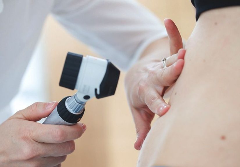
2) Dermoscopy
Dermatologists often use a handheld device called a dermatoscope (dermoscopy) to see structures beneath the surface. This can help distinguish
benign growth patterns from suspicious ones.
3) Biopsy (the “know for sure” step)
If a spot is suspiciousor if it’s unclearyour clinician may recommend a skin biopsy. A biopsy removes a small piece (or all)
of the lesion so a lab can examine it under a microscope. This is the standard way to confirm whether a lesion is melanoma or something benign.
Many biopsies are quick, done with local numbing medicine, and take just minutes. The goal isn’t to be dramaticit’s to be precise.
Treatment Differences (Because the Plan Depends on the Diagnosis)
If it’s seborrheic keratosis
SKs often don’t need treatment. Removal may be offered if it’s irritating, itchy, frequently snagging, or cosmetically bothersome.
Common in-office removal options may include:
- Cryotherapy: freezing the lesion
- Curettage: gently scraping the growth off
- Shave removal: shaving off the raised portion
- Electrosurgery: using controlled electrical energy (often paired with curettage)
Your clinician will choose a method based on the lesion’s size, location, and appearanceand whether the spot needs biopsy first.
If it’s melanoma
Melanoma treatment depends on how deep the tumor is and whether it has spread. Early melanoma is often treated with surgical removal
(wide local excision). For higher-risk cases, clinicians may evaluate lymph nodes (for example, sentinel lymph node biopsy) and consider
additional therapies. The important takeaway for this article: early detection is a big deal.
Risk Factors and Prevention You Can Actually Use
Melanoma risk factors (common ones)
- History of intense sun exposure or sunburns
- Indoor tanning
- Many moles or atypical moles
- Personal or family history of melanoma
- Weakened immune system
Sun safety basics (the boring advice that works)
Sun protection isn’t about never seeing daylight againit’s about reducing UV damage over time.
- Seek shade when possible, especially during peak sun hours
- Wear protective clothing, a wide-brim hat, and UV-blocking sunglasses
- Use broad-spectrum sunscreen and reapply as directed, especially after swimming or sweating
- Avoid indoor tanning (your future skin will thank you)
How to do a monthly skin self-check
- Use good lighting and a full-length mirror, plus a hand mirror for hard-to-see areas.
- Check front and back of your body, then sides, arms, underarms, hands, and nails.
- Look at legs, feet, soles, and between toes.
- Don’t forget scalp and behind ears (a comb or a helper can be useful).
- Note anything new or changing; take a clear photo with a date for comparison.
Photos can help you track change, but they’re not a diagnosis. If something is evolving, get it checked.
FAQs People Ask (Usually While Staring at a Spot in the Mirror)
Can a seborrheic keratosis turn into melanoma?
Seborrheic keratosis is considered benign and typically does not “transform” into melanoma. The bigger issue is that some SKs can
resemble melanoma or sit near other lesionsso any suspicious change still deserves evaluation.
Can melanoma look like a scab or wart?
Melanoma can sometimes crust, bleed, or look like an irritated spot. And some melanomas don’t look like the textbook “mole with jagged edges.”
If it’s persistent, changing, or repeatedly bleeding, don’t assume it’s just a scab.
What if my spot is raised and itchy?
Itchiness can happen with benign lesions (including SKs), irritation, eczema, and more. It can also occur with skin cancers.
Itch alone isn’t diagnosticthe pattern over time matters.
Should I remove SKs at home?
It’s safer to avoid at-home removal attempts. First, you want to be sure it’s truly benign. Second, DIY removal can lead to infection,
scarring, and confusion if the lesion changes afterward. If it bothers you, a clinician can remove it properly (and decide if biopsy is needed).
The Bottom Line: A Simple Rule That Covers 99% of Real Life
If a spot is new, changing, bleeding, crusting, painful, or just looks different from the rest of your skin marks,
it’s worth a professional look. Many “scary-looking” lesions are benign, and many early cancers are subtleso when in doubt, check it out.
Think of a dermatologist visit like a seatbelt: you hope you never “need” it, but you’ll be glad it exists.
: experiences section
Real-World Experiences: What People Commonly Go Through When Comparing SK vs. Melanoma
If you’ve ever zoomed in on a skin spot with your phone camera until it looks like a satellite photo of Mars, you’re in very good company.
One of the most common experiences people report is the emotional whiplash of noticing a new growth and immediately imagining worst-case scenarios.
That reaction is understandable: skin changes are visible, and “visible” often feels “urgent.” The challenge is that skin is also full of harmless
surprisesespecially as we ageso panic and reality don’t always match.
1) The “It appeared out of nowhere!” moment
A frequent story goes like this: someone spots a dark, raised patch on the back, shoulder, or chest while changing clothes or stepping out of the
shower. Because seborrheic keratoses can start as small bumps and gradually thicken, it can feel like they popped up overnightwhen in fact they
may have been slowly developing for months. The surprise factor alone can be scary.
2) The “Google spiral” (and why it’s so common)
People often try to compare photos online and end up more confused. That’s because SKs and melanoma can overlap in appearanceespecially when an SK
is very dark, irritated, or crusty. Many people describe feeling relieved when they learn that “stuck-on” and waxy texture points toward SKbut also
surprised to hear that visual cues aren’t enough for certainty. In other words: online images can help you notice patterns, but they can’t diagnose you.
3) The dermatologist visit that turns anxiety into clarity
Another common experience is realizing how fast a professional exam can change the whole situation. Dermatologists often recognize classic SKs quickly,
sometimes using dermoscopy to confirm the pattern. People frequently report feeling immediate relief when the clinician says, “This looks benign.”
On the flip side, if a lesion looks suspicious, patients often appreciate that the next step is concrete: biopsy and answersrather than endless guessing.
4) The biopsy experience is usually less dramatic than expected
Many people imagine a biopsy as a big procedure. In reality, the typical experience described is: numbing medicine, quick removal of a small sample,
a bandage, and some basic wound care instructions. The waiting period for results can feel like the hardest part, but people often say that having a plan
(and a clear timeline for follow-up) helps them cope.
5) The “I’m glad I went in early” lesson
Perhaps the most consistent takeaway across real-world stories is this: people are grateful when they didn’t ignore change. Sometimes the result is
“just an SK,” and that’s a winbecause the worry ends and the irritation can be treated. Other times, early evaluation catches melanoma at a stage where
treatment is more straightforward. Either way, the experience reinforces a practical mindset: you don’t need to be 100% sure it’s dangerous to get it checked.
You only need to be 1% unsureand willing to choose certainty over stress.
If you’re currently in the “Is this SK or melanoma?” headspace, you’re not overreactingyou’re paying attention. The goal isn’t to become your own dermatologist.
The goal is to notice change, respect the warning signs, and let a qualified professional confirm what’s going on.