Table of Contents >> Show >> Hide
- What a Breast Ultrasound Is (and What It Isn’t)
- Purpose: Why Providers Order an Ecografía Mamaria
- How the Test Works (A Quick “Science Without the Sigh” Explanation)
- How to Prepare (So You Don’t Arrive Wearing the Wrong Things)
- The Procedure: What Actually Happens in the Room
- Types of Breast Ultrasound You Might Hear About
- Results: How They’re Reported and What They Mean
- Limitations and Tradeoffs (Because Every Test Has Them)
- Practical Questions to Ask Your Clinician (Yes, You’re Allowed)
- A Calm, Honest Bottom Line
- Real-World Experiences: What the Appointment Feels Like (and What People Commonly Notice)
- SEO Metadata (JSON)
“Ecografía mamaria” is simply the Spanish way to say breast ultrasound (also called a breast sonogram).
If you’ve been scheduled for one, you might be picturing something dramaticsirens, suspense music, a doctor whispering, “Zoom in.”
In real life, it’s usually much less cinematic and much more: gel, a warm room, and a wand that looks like a TV remote.
Quick, painless for most people, and genuinely usefulespecially when your provider wants a closer look at something in the breast.
This guide breaks down what a breast ultrasound is for, what happens during the exam, and how to interpret the results (including BI-RADS language).
You’ll also get practical tips to feel more preparedbecause “show up and wing it” is not a vibe when you’re already anxious.
What a Breast Ultrasound Is (and What It Isn’t)
A breast ultrasound uses sound waves to create images of breast tissue. Unlike a mammogram, it does
not use X-rays. The ultrasound machine sends sound waves into the tissue and listens for the echoes to build pictures on a screen.
Think of it as “bat sonar,” but for radiologyminus the cape.
Here’s the key distinction: a breast ultrasound is most often a diagnostic tool (used to investigate a specific concern),
not a universal replacement for mammograms. Mammography is still the primary screening test for most people because it images the entire breast
and can detect certain findings (like calcifications) that ultrasound may not show as well.
Purpose: Why Providers Order an Ecografía Mamaria
Breast ultrasound is commonly ordered when your healthcare team wants more information than a physical examor even a mammogramcan provide.
It’s especially helpful for sorting out what’s happening in a focused area.
1) To evaluate a lump or new breast change
If you or your provider can feel a lump, ultrasound can help determine whether it looks like a
fluid-filled cyst or a solid mass. That difference matters because simple cysts are usually benign,
while solid masses often need additional evaluation.
2) To take a closer look after an abnormal or unclear mammogram
Sometimes a mammogram shows an area that needs a “second camera angle.” Ultrasound can zoom in on a specific region to clarify what’s going on,
especially if the mammogram result is incomplete or the tissue is dense.
3) As supplemental imaging in people with dense breast tissue
Dense breast tissue can make mammograms harder to read because dense tissue and some abnormalities can both appear white on the image.
In some cases, clinicians may discuss ultrasound (or MRI) as additional imagingparticularly when individual risk factors suggest it may be helpful.
However, experts don’t universally agree on which additional tests should be routine for everyone with dense breasts, because extra imaging can also
increase “false alarms” and lead to more tests and biopsies that ultimately aren’t cancer.
4) To guide a needle for biopsy or fluid aspiration
Ultrasound isn’t just for picturesit can also be used in real time to guide a needle to the right spot during
an ultrasound-guided biopsy or when draining a painful cyst. This helps clinicians sample the tissue (or fluid)
from exactly where they need it.
5) To evaluate symptoms like nipple discharge or underarm lymph nodes
Depending on symptoms, ultrasound may be used to investigate concerns such as clear or bloody nipple discharge, focal breast pain,
or areas in the armpit where lymph nodes live (and sometimes misbehave).
How the Test Works (A Quick “Science Without the Sigh” Explanation)
During the exam, a technologist (sonographer) applies a clear gel to the skin and moves a handheld device called a
transducer across the breast. The transducer sends sound waves into the tissue and collects the returning echoes,
which the computer converts into live images.
Sometimes a Doppler ultrasound is used to look at blood flow patterns within a region of interest.
If Doppler is used, you might hear pulsing sounds that change as blood flow is measuredtotally normal, mildly “spaceship,” and not a cause for panic.
How to Prepare (So You Don’t Arrive Wearing the Wrong Things)
- Skip deodorant, lotions, or powders on or around the breasts and underarms that day. These can interfere with clear imaging.
- Wear a two-piece outfit if possible. You’ll undress from the waist up and change into a front-opening gown.
- Bring prior imaging info if you had mammograms or ultrasounds at another facility. Comparisons over time can be very helpful.
- Know your timeline: Ask whether you’ll get results the same day or through your patient portal later.
- Mention relevant context: pregnancy, breastfeeding, implants, recent surgery, or a known lump location.
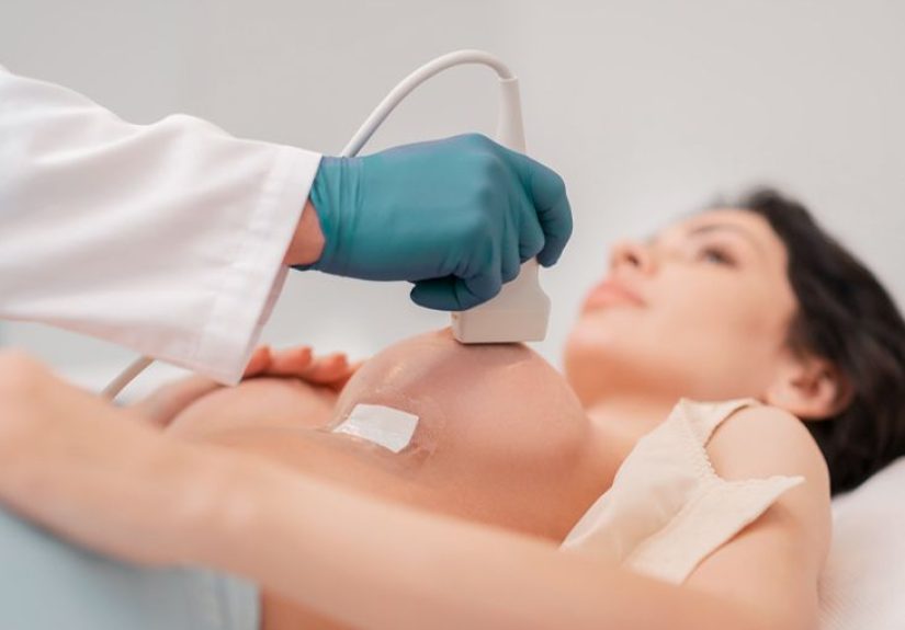
The Procedure: What Actually Happens in the Room
Most breast ultrasounds take about 30 minutes, and you can typically return to normal activities immediately afterward.
Here’s a realistic play-by-play:
- Check-in and gown time: You’ll undress from the waist up and put on a gown that opens in front.
- Positioning: You’ll lie on an exam table, often with your arm above your head to help flatten and expose the area being scanned.
- The gel: A clear gel is applied to improve contact and help sound waves travel. It can feel cool. No one loves it. We all survive it.
- Scanning: The transducer is moved across the breast and sometimes the underarm. You may be asked to roll slightly side to side.
- Pressure notes: Usually it’s not painful, but tender areas can feel pressure or mild discomfort.
- Image review: The technologist captures images, and a radiologist later interprets them (sometimes the radiologist reviews while you wait).
Pro tip: If the technologist gets quiet, it doesn’t automatically mean “bad news.” It can mean “I’m concentrating,” “I need one more angle,”
or “I’m making sure the images are crisp enough that the radiologist doesn’t send me back in there.” Silence is not a diagnosis.
Types of Breast Ultrasound You Might Hear About
Handheld diagnostic ultrasound
This is the most common typeperformed with a handheld transducer over the region of interest.
Automated Breast Ultrasound (ABUS)
Some centers use automated systems (often discussed for dense breasts) that scan more of the breast in a standardized way.
Availability varies, and whether it makes sense depends on your individual risk and your clinician’s plan.
Ultrasound-guided procedures
If imaging suggests a biopsy or aspiration is appropriate, ultrasound may be used during the procedure to guide the needle to the target.
This is commonly done with local numbing medication and tends to be less invasive than surgical biopsy.
Results: How They’re Reported and What They Mean
After the exam, a radiologist (a physician trained to interpret imaging) reviews the images and sends a report to the clinician who ordered the test.
How quickly you hear back depends on the facility and whether the ultrasound was urgent, but many imaging centers provide results quicklysometimes even the same day.
Common “normal” or benign findings
Ultrasound can show many non-cancerous (benign) conditions. Examples often mentioned in reports include:
- Simple cysts: fluid-filled sacs that are usually benign
- Fibroadenomas: common benign solid growths
- Lipomas: benign fatty lumps
Suspicious findings and the BI-RADS system
Many breast imaging reports use BI-RADS (Breast Imaging Reporting and Data System), a standardized scoring framework.
BI-RADS is used for mammograms, breast ultrasound, and breast MRI. It helps your care team communicate clearly about the level of concern and next steps.
The exact wording differs by facility, but BI-RADS categories typically include:
- BI-RADS 0: Incompletemore imaging needed
- BI-RADS 1: Negative (no concerning findings)
- BI-RADS 2: Benign finding (routine screening as appropriate)
- BI-RADS 3: Probably benignshort-interval follow-up imaging often recommended
- BI-RADS 4: Suspiciousbiopsy may be recommended (often subdivided by level of suspicion)
- BI-RADS 5: Highly suggestive of malignancybiopsy strongly recommended
- BI-RADS 6: Known biopsy-proven malignancy (used after cancer is confirmed)
What happens next depends on the category
Here are realistic next-step scenarios:
- “It’s a simple cyst”: Often no treatment is needed unless it’s painful or problematic; sometimes aspiration is offered for comfort.
- “Probably benign” (BI-RADS 3): Follow-up imaging may be suggested to confirm stability over time.
- “Suspicious” (BI-RADS 4/5): Your team may recommend a biopsy to determine what the cells actually are (imaging can’t always tell you for sure).
- “Needs correlation”: The radiologist may recommend comparing with prior images or adding another modality (mammogram, MRI) for completeness.
Important note: An ultrasound result that leads to a biopsy recommendation is not the same as a cancer diagnosis.
Biopsies are frequently done out of caution, and many come back benign.
Limitations and Tradeoffs (Because Every Test Has Them)
A breast ultrasound is safe and doesn’t expose you to ionizing radiation. But it has limits, and knowing them can reduce anxiety and prevent unrealistic expectations.
It’s not a perfect “all-in-one” screening tool
Ultrasound may not show certain breast changes as well as mammography doescalcifications are a classic example.
That’s one reason mammography remains a cornerstone of screening.
More imaging can mean more follow-ups
Supplemental imaging (like ultrasound in dense breasts) can help find some cancers not seen on mammograms, but it can also identify findings that aren’t cancer.
That can lead to extra imaging, short-interval follow-ups, or biopsies that ultimately show benign results.
Whether that tradeoff is worth it depends on your personal risk factors and your values (some people prefer maximum sensitivity, others prefer fewer callbacks).
“Dense breasts” doesn’t automatically mean “you need everything”
Dense breast tissue is common, and it can slightly raise breast cancer risk while also making mammograms harder to interpret.
However, major experts have noted that evidence is not definitive enough to recommend for or against supplemental imaging for all women with dense breasts.
That’s why the best plan is personalizedbased on family history, genetics, prior biopsies, age, and overall risk assessment.
Practical Questions to Ask Your Clinician (Yes, You’re Allowed)
- What was my BI-RADS category, and what does it mean for me?
- Is the finding a simple cyst, a complex cyst, or a solid mass?
- Do I need follow-up imaging? If so, when and why that interval?
- If a biopsy is recommended, will it be ultrasound-guided?
- Do my breast density and risk factors suggest any changes to my screening plan?
- When and how will I receive the written report?
A Calm, Honest Bottom Line
An ecografía mamaria (breast ultrasound) is one of the most common, useful follow-up tests in breast imaging.
It’s quick, doesn’t use radiation, and can clarify whether a lump looks like a simple cyst or a solid mass.
It can also guide biopsies when sampling is needed.
The results can feel like a new languageespecially when BI-RADS enters the chatbut the goal is simple:
sort benign from “needs a closer look,” and make sure nothing important is missed.
If you walk away with one takeaway, let it be this: an ultrasound is information, not a verdict.
And you deserve clear explanations for whatever the next step is.
Real-World Experiences: What the Appointment Feels Like (and What People Commonly Notice)
The medical description of a breast ultrasound is accurate, but it’s often missing the human detailsthe parts people actually remember.
Below are common experiences patients report, shared here as composite stories (not medical advice, and not a substitute for talking with your clinician).
The goal is to make the process feel familiar before you’re lying on a table wondering whether the gel is supposed to be that cold. (It is.)
The “Callback” Spiral
One of the most common paths to ultrasound is a callback after a mammogram. Many people describe getting that message and immediately imagining the worst,
even though callbacks are common and often lead to benign explanations. In the appointment, the anxiety usually peaks during the moments of silence
when the technologist is capturing images and not narrating every click. What helps: reminding yourself that the technologist’s job is image quality,
not interpretation, and that “quiet” is often just “focused.” People often feel a wave of relief simply because the ultrasound provides more clarity:
“This looks like a cyst” lands very differently than “We’re not sure what we’re seeing.”
The “I Found a Lump in the Shower” Moment
Another common experience: noticing a new lump and feeling that immediate, surreal mix of disbelief and urgency.
People often worry they’ll be judged for waiting a few days, but clinicians expect thislife happens, and most lumps are benign.
Ultrasound appointments for palpable lumps tend to feel straightforward: point to the spot, lie down, scan that region carefully.
Patients often say the most surprising part is how normal the room feelsbright lights, a screen, a calm routinewhile their brain is doing a full
disaster montage. When the radiologist later explains what was seen (for example, a simple cyst or a likely fibroadenoma), the emotional whiplash can be real:
you walk in braced for bad news and walk out with a plan, which is sometimes the most valuable “result” of all.
Dense Breasts and the “Wait…Am I High Risk?” Question
People with dense breasts often describe confusion after reading their mammogram notification: “Does this mean they can’t see anything?” or
“Does this mean I’m going to get cancer?” The honest answer is more nuanced. Many patients feel better after a clinician explains that dense tissue is common,
that mammograms still matter, and that extra imaging is a risk-and-benefit conversationnot an automatic upgrade package.
Some people appreciate the additional reassurance of supplemental ultrasound; others find that extra testing increases anxiety due to follow-up imaging.
The shared experience is wanting a clear, personalized recommendation instead of generic “you might consider…” language.
The Biopsy-Adjacent Experience
When an ultrasound leads to a biopsy recommendation, patients commonly report feeling like the floor drops outuntil someone explains what that recommendation
actually means: imaging can identify features that might be concerning, but only tissue can confirm the diagnosis. Many people say the best moment
in the process is when the plan becomes concrete: an ultrasound-guided biopsy date, a clear outline of what will happen, and a realistic timeframe for results.
Even if the waiting is stressful, having a defined next step can feel more manageable than uncertainty.
If you’re in this situation, it can help to ask: “What is the BI-RADS category?” and “What specific feature made biopsy the best next step?”
Clarity doesn’t erase anxiety, but it does turn the anxiety from fog into something you can navigate.
Small Comforts People Swear By
A few practical things people mention again and again: wear a soft, easy-to-remove top; bring a hair tie; don’t schedule a high-stakes meeting immediately
afterward if you can avoid it; and plan a small “after” reward (coffee, a walk, texting a friend) because your nervous system will have earned it.
The ultrasound itself is usually quickwhat takes time is the emotional build-up beforehand.
Many patients leave feeling surprised by how routine the test was, and grateful they didn’t have to sit with uncertainty for weeks.